The medial collateral ligament’s main function is to prevent the leg from extending too far inward, but it also helps keep the knee stable and allows it to rotate. Injuries to the medial collateral ligament most often happen when the knee is hit directly on its outer side.
What is the function of the tibial collateral ligament?
The knee relies on ligaments, which connect bone to bone, and surrounding muscles for stability. The primary function of the tibial collateral ligament is to provide additional stability to the knee joint itself.
What movement does the ACL prevent?
The cruciate ligaments control the back and forth motion of your knee. The anterior cruciate ligament runs diagonally in the middle of the knee. It prevents the tibia from sliding out in front of the femur, as well as provides rotational stability to the knee. Normal knee anatomy.
What is the function of the tibial and fibular collateral ligaments?
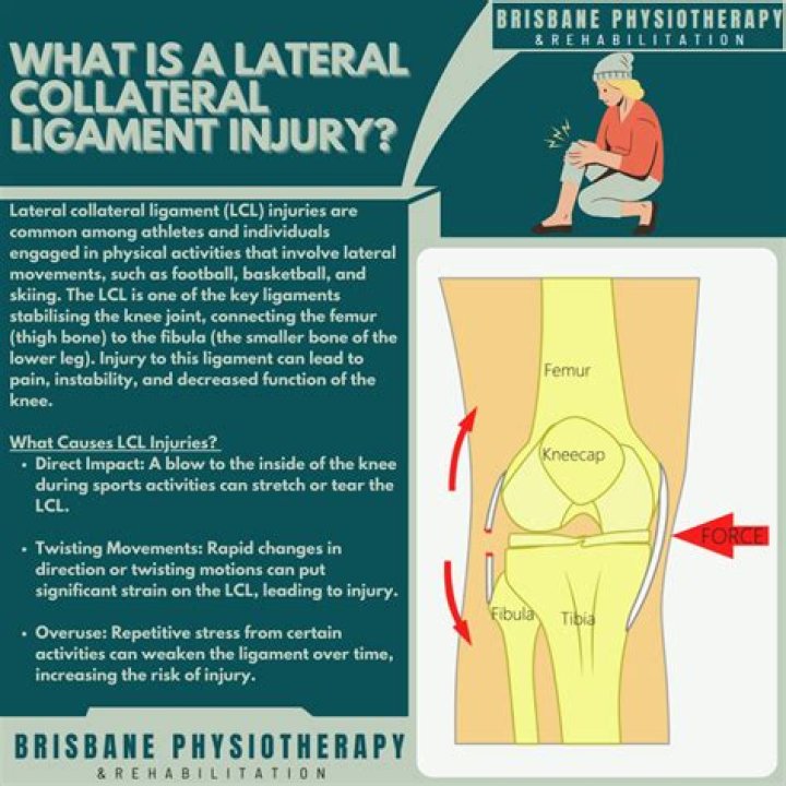
Collateral ligaments. They control the side to side motion of your knee and brace it against unusual movement. The medial collateral ligament (MCL) is on the inside. It connects the femur to the tibia. The lateral collateral ligament (LCL) is on the outside.What motion does the lateral collateral ligament prevent?
Lateral collateral ligament (LCL) – prevents medial movement of the tibia on the femur when varus (towards the midline) stress is placed on the knee. Runs between the lateral epicondyle of the femur and the head of the fibula. Also known as the fibular collateral ligament (FCL).
What does MCL limit?
An MCL is the legal threshold limit on the amount of a substance that is allowed in public water systems under the Safe Drinking Water Act (SDWA). The limit is usually expressed as a concentration in milligrams or micrograms per liter of water.
What does the deltoid ligament prevent?
Deltoid ligament functions To prevent the talus shifting into a valgus position, or to move anterolaterally, or to externally rotate. To transfer force between the tibia and tarsus.
What movements does the fibular collateral ligament prevent?
The lateral collateral ligament is located on the outside of the knee joint, and it connects your femur to your fibula (a lower-leg bone that is smaller than the tibia). 3 The LCL prevents excessive adduction of the knee (i.e., movement toward the central axis of the body).Why are ligaments important in the knee?
Tendons connect the knee bones to the leg muscles that move the knee joint. Ligaments join the knee bones and provide stability to the knee: The anterior cruciate ligament prevents the femur from sliding backward on the tibia (or the tibia sliding forward on the femur).
Why are popliteal ligaments important?Its most important role is providing forward stabilization of the knee, and also the stabilization of the retraction of the backside of the knee joint and the lateral meniscus during the flexion of the knee. It allows the knee to flex when it is in full extension.
Article first time published onWhat does the ACL stabilize?
The anterior cruciate ligament (ACL) is one of the key ligaments that help stabilize your knee joint. The ACL connects your thighbone (femur) to your shinbone (tibia). It’s most commonly torn during sports that involve sudden stops and changes in direction — such as basketball, soccer, tennis and volleyball.
What muscles protect the ACL?
Quadriceps muscle contrac- tion protects the anterior cruciate ligament during anterior tibia1 translation.
Which knee ligament prevents posterior movement of the tibia?
The function of the PCL is to prevent the femur from sliding off the anterior edge of the tibia and to prevent the tibia from displacing posterior to the femur. The posterior cruciate ligament is located within the knee.
Which ligament prevents abduction of the tibia?
Your medial collateral ligament prevents excessive abduction of the tibia and guards an excessive force coming from the outside area of your knee. Your lateral collateral ligament prevents excessive adduction of the tibia and guards against an excessive force coming from the inside aspect of your knee.
What are collateral ligaments?
A ligament is a band of tissue that connects a bone to another bone. The collateral ligaments of the knee are located on the outside part of your knee joint. They help connect the bones of your upper and lower leg, around your knee joint. The lateral collateral ligament (LCL) runs on the outer side of your knee.
What structures prevent the excessive lateral and medial movements of the tibia at the knee?
medial collateral ligament (MCL), which gives stability to the inner part of the knee. lateral collateral ligament (LCL), which stabilizes the outer part of the knee. anterior cruciate ligament (ACL), which is located in the center of the knee and prevents excessive forward movement of the tibia.
What bone articulates with the distal tibia?
Distally, the tibia articulates with the talus to form the talocrural joint of the ankle. Although not functionally a part of the knee, the fibula articulates proximally with the lateral aspect of the tibia, forming the proximal tibiofibular joint.
How do I strengthen my medial ankle ligaments?
- Trace the alphabet with your toe, which encourages ankle movement in all directions. Trace the alphabet 1 to 3 times.
- Sit in a chair with your foot flat on the floor. Slowly move your knee side to side while keeping your foot pressed flat. Continue for 2 to 3 minutes.
What ligaments attach the tibia to the fibula?
anterior tibiofibular ligament, which connects the tibia to the fibula.
What muscles stabilize the MCL?
While the MCL is the static stabilizer of the medial knee, the dynamic stabilizers of the medial knee are muscles: the semimembranosus complex, vastus medialis, and pes anserinus.
How important is the MCL?
The medial collateral ligament is one of four major ligaments that support the knee. The MCL is the prime static stabilizer of the medial side of the knee joint, and is important for providing support against valgus stress, rotational forces, and anterior translational forces on the tibia.
What muscles attach to MCL?
The medial collateral ligament, which is also known as the tibial collateral ligament, is a broad, flat, bandlike ligament that runs from the medial condyle of the femur to the medial aspect of the shaft of the tibia, where it attaches just above the groove where the semimembranosus muscle attaches (Fig. 107.2).
How do you strengthen knee ligaments?
- Benefits.
- Leg lifts.
- Standing hamstring curls.
- Hamstring curls on a weight bench.
- Step exercises.
- Single-leg dip.
- Wall squats.
- Post-exercise stretching.
What muscles Stabilise knees?
The muscles surrounding the knee function to both move and stabilize the joint. The two main muscle groups are the quadriceps on the anterior side of the knee and femur, and the hamstrings on the posterior side.
What type of movement does the ACL prevent in the knee?
The ACL prevents the tibia from sliding too far forward from underneath the femur. It also helps prevent hyperextension of the knee, and resistance to rotational forces about the knee. ACL knee surgery is necessary once the tibia has slid too far or the knee joint has experienced hyperextension.
Which ligaments restrict movement of femur on tibia?
The ACL prevents the femur from sliding backwards on the tibia (or the tibia sliding forwards on the femur). Together with the posterior cruciate ligament (PCL), ACL stabilizes the knee in a rotational fashion.
What attaches to the tibial tuberosity?
Structure. The tuberosity of the tibia gives attachment to the patellar ligament, which attaches to the patella from where the suprapatellar ligament forms the distal tendon of the quadriceps femoris muscles. The quadriceps muscles consist of the rectus femoris, vastus lateralis, vastus medialis, and vastus intermedius …
What muscle gives rise to the oblique popliteal ligament?
It is one of the five insertions of the semimembranosus muscle. The oblique popliteal ligament forms part of the floor of the popliteal fossa, and the popliteal artery rests upon it. It is formed of fasciculi separated from one another by apertures for the passage of vessels and nerves.
Which of the following provides the most stability to the elbow?
The important ligaments of the elbow are the medial collateral ligament (on the inside of the elbow) and the lateral collateral ligament (on the outside of the elbow.) Together these ligaments provide the main source of stability for the elbow, holding the humerus and the ulna tightly together.
What is the function of the Arcuate and popliteal ligaments of the knee?
The posterior cruciate ligament and the arcuate ligament have predominant role for the posterolateral stability of the knee. The functional restoration of these ligaments is an important part of the surgical treatment of posterolateral ligamentous injuries.
Does ACL connect tibia to femur?
The anterior cruciate ligament (ACL) connects the femur to the tibia and stabilize the knee join. The ACL resists anterior translation of the tibia relative to the femur; it also resists rotation.
